By Mary Sanderson
The neck flexors are generally weaker than the extensors, which have to hold the heavy head in an upright position against gravity. The extensors are constantly under tension, contracting statically and eccentrically to maintain posture.
Postural deficiencies can especially occur with repetitive activities or positions, such as sitting, writing for long periods, painting a ceiling, or engaging in sports like cycling. Soft tissues can become micro-torn and tense; as the activity continues, holding patterns and imbalances prevail. An increase in the cervical curve, forcing the head forwards, is a common result.
Problems manifest as movement restrictions, headaches, vertigo, tinnitus, and muscle, joint and nerve pain. Vertebral artery and nerve impingement can occur, which may not have a muscular cause, and medical advice should be sought if a subject presents with dizziness or referred pain that is not clearly muscular in origin.
As well as controlling specific movements, many of the small neck muscles are involved in maintaining the balance and stability of the head on the neck; these cannot be palpated, so they will not be discussed. The platysma is the most superficial anterior muscle and is a thin, flat muscle that adheres to the skin.
Seated STR is a good way to start assessment and treatment, and you can generally address the neck flexors if the head and neck are protracted. Then, systematically working the agonists and antagonists will support recovery from chronic neck tension and side effects such as tension-type headaches (TTHs). This will also support a return to good posture and improved functional capacity.
Because of its extreme mobility, the neck is also vulnerable to traumatic injury, an example being whiplash. After such an injury, ligamentous damage occurs, and the neck muscles become extremely tense. This is due to fierce reflex muscle contractions that protect the head against rapid movement. If medical screening results are satisfactory, STR is an indispensable therapy. Degenerative conditions, such as spondylosis, can also benefit from STR; active STR ensures the subject moves only through a range that can be performed comfortably. Improving movement and posture will relieve pressure on the facet joints and discs
|
Muscle |
Effects of muscle restrictions |
|
Suboccipitals • Rectus capitis posterior minor • Rectus capitis posterior major • Superior oblique • Inferior oblique |
Limited studies exist on the effects of tightness in these muscles, although connections to dura are found in rectus capitis posterior minor, which is associated with headaches. There is also a suggestion that tightness may affect positioning of head, increasing upper cervical extension. |
|
Semispinalis capitis |
Reduced flexion of head and cervical spine. Entrapment of greater occipital nerve. |
|
Splenius capitis Splenius cervicis |
Reduced flexion of head and cervical spine. Limited studies exist on these muscles. |
|
Longissimus capitis |
Increased lateral flexion of head on neck when observed in frontal plane, and posterior rotation of cervical spine. |
|
Levator scapulae |
Pain in medial scapula and neck. Increased ipsilateral neck flexion and/or raised scapula. |
|
Trapezius |
Reduced head and cervical spine flexion and ipsilateral rotation of head. Head pain is thought to be linked to trigger points in tight upper fibres. |
|
Sternocleidomastoid |
Torticollis, both spasmodic and congenital. Head-forward posture. Increased extension of upper cervical spine. |
|
Scalenes |
Sensation changes in arms and hands as the brachial plexus is compressed. Reduced rotation and increased lateral flexion of cervical spine. |
Neck Flexion
Major Muscles: SCM, scalenus anterior and longus colli (flex the neck); longus capitis and SCM (flex the neck and head); rectus capitis anterior (flexes the head on the neck and stabilises the atlanto-occipital joint).
Neck Side Flexion
Major Muscles: Scalenes, splenius cervicis, levator scapulae and SCM (side flex the neck); SCM, splenius capitis, trapezius and erector spinae (side flex the head and neck); rectus capitis lateralis (side flexes the head on the neck).
Neck flexion occurs when the SCM muscles on both sides contract; side flexion to the same side or rotation to the opposite side occurs when the SCM on one side contracts. When the head and neck are fixed, the SCM muscles can raise the clavicles and sternum, thus assisting inspiration. The scalenes, when contracting bilaterally, also assist in neck flexion; if only one side contracts, they assist in side flexion to the same side. The brachial plexus runs between the scalenus anterior and the scalenus medius.
Neck Flexors and Side Flexors –
Treatment
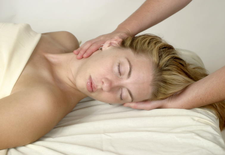
With the subject in a supine position, support the head with one hand and gently grasp the SCM with the other hand. Maintain this hold and instruct the subject to extend the neck.
Alternatively, gently grasp the muscle and passively move the neck away from this lock into side flexion; or instruct the subject to rotate the neck to the same side for a stretch. Do not move too quickly. If the area is particularly congested, apply pressure to one side of the muscle at a time. Lock into the muscle origin, moving to two or three new points to free the clavicular and sternal fibres. STR at the SCM insertion points on the skull is necessary, and an MFR lock will prove highly successful where fascial thickening is often evident; using the knuckle away from the bone helps attain a lock, but move carefully into position.
The scalenes assist with inspiration, so incorporating breathing will not only help to relax the subject, but also directly assist with the release. You can treat the scalenes by first side-flexing the neck to the same side during inhalation, then gliding away from the clavicle with the lock and moving the head into side flexion to the opposite side during exhalation. For scalenus anterior, lock in just laterally to the SCM; for the scalenus medius, lock in away from the mid-portion of the clavicle; and for the scalenus posterior, lock in away from the most lateral point of the clavicle.
Because this whole area can be highly sensitive, treatment should always be slow.
Neck Extension
Major Muscles: levator scapulae and splenius cervicis (extend the neck); trapezius, splenius capitis, and erector spinae (extend the head and neck); rectus capitis posterior major and minor, and superior oblique (extend the head on the neck).
Neck Rotation
Major Muscles: Semispinalis cervicis, multifidus, scalenus anterior and splenius cervicis (rotate the neck); splenius capitis and SCM (rotate the head and the neck); inferior oblique and rectus capitis posterior major (rotate the head on the neck).
Neck Extensors and Rotators – Treatment
There are two main ways to work this area. Firstly, with the subject in a supine position, support the head with one hand and lock into the extensor muscles with the other hand as the neck is flexed, side flexed or rotated. Systematically release the superficial layers of the entire back and side of the neck; often, only minimal movement is required for an effective result.
Congestion often occurs between the trapezius and the SCM, within the splenius muscles, and in the levator scapulae. These muscles may be reached by locking in deep to the lateral border of the SCM as the neck is side-flexed to the opposite side, or as the subject is instructed to flex the head.
The upper fibres of the trapezius can be addressed using an index finger reinforced with the middle finger as the neck is flexed or side flexed to the opposite side. The deeper extensors can be engaged as the chin is tucked in for a stretch.
Use an MFR lock to engage the suboccipital muscles as the subject is instructed to tuck the chin in.
Second, in a seated position, the subject can perform a variety of side flexion, flexion, and rotation movements as the therapist provides a specific lock. This is a highly effective way of introducing functional awareness as the subject is guided into a stretch.
Apply pressure gently and precisely, or movement will be difficult. This approach can treat the trapezius and levator scapulae highly effectively. You can target the levator scapulae insertion point by curling under the anterior fibres of the trapezius toward the medial border of the scapula; the subject can then side-flex and flex the neck.
Temporomandibular Joint (TMJ)
Four main muscles are associated with this joint: the temporalis, masseter, and the medial and lateral pterygoids.
Problems present as a general aching in the area, restricted or asymmetrical movement, and clicking. Dysfunction can occur due to trauma, for example, a whiplash injury or a direct blow in a contact sport. Tissue congestion can develop from a jaw fracture or tooth loss, or after major dental surgery during which the mouth has been forced open for long periods. Chewing on one side of the mouth or clenching the teeth may cause an overuse injury, which can lead to pain and headaches.
TMJ – Treatment
If you have problems in this area, you may need specialist advice, particularly from a dentist, who will check your bite.
STR treatment should initially include general treatment for the neck. STR to the muscles that move the joint will help reduce pain and support re-education of faulty movement patterns; often, a dentist will suggest exercises, such as placing the tongue on the upper palate as the mandible opens.
|
Muscle |
Effects of muscle restrictions |
|
Masseter |
Restricted jaw depression, lateral deviation of mandible (unilateral) with impact on bite mechanics. Compression of disc and tissue. |
|
Temporalis |
Similar to those of masseter but with greater effect. Static retraction of jaw and concomitant reduction in mandible protraction. Linked to headaches and pain. |
|
Lateral pterygoid |
Subluxation of mandible and dysfunctional movement between disc and mandible, resulting in clicks and possible locking of jaw. Linked to headaches. |
|
Medial pterygoid |
Compression of disc and if unilateral, an increased contralateral deviation of jaw, affecting bite mechanics. |
Apply an MFR lock to the temporalis and ask the subject to open the mouth.
Then apply a lock to the masseter and again ask the subject to open the mouth. Reinforce the index finger with the middle finger to work more deeply and closer to the TMJ to target the pterygoids. Initially, you can treat both sides at the same time; observe how wide the mouth opens and note any deviation while palpating the tissues. For more specific STR, perform the lock to one side at a time, but avoid over-treating.